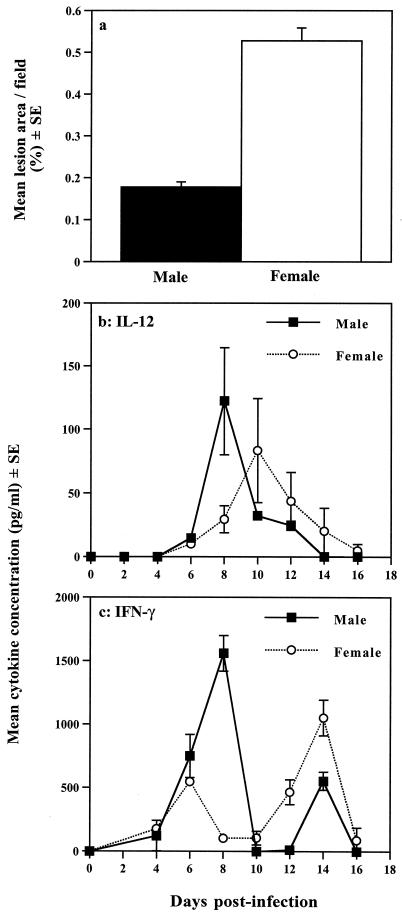
FIG. 2.
Innate resistance to T. gondii is influenced by gender. (a) Determination of the area of brain lesions in T. gondii-infected male and female SCID mice. Chalkley counting (a microscpic method of determining area) of histological sections of brain tissue derived from animals at day 15 postinfection revealed that brains from female animals contained significantly greater lesion areas than those of their male counterparts. Female lesion area, 0.527%; male lesion area, 0.177%; P < 0.01. (b and c) Comparison of innate immunity-associated cytokines in male and female SCID mice infected with T. gondii. (b) Plasma samples from infected animals analyzed for IL-12 expression revealed significant differences between males and females. IL-12 plasma concentrations peaked at day 8 postinfection in males (122.3 ± 42.2 pg/ml) and day 10 in females (83.3 ± 40.8 pg/ml). The level of IL-12 in male samples was significantly higher at day 8 postinfection compared with females (P < 0.03). (c) Similar measurements of IFN-γ levels revealed that male SCID mice produced significantly higher amounts of this cytokine early after infection than females. At day 8, male IFN-γ plasma levels were 1,560 ± 140 pg/ml, compared with 104 ± 10.4 pg/ml in females (P < 0.001). This pattern of cytokine expression was consistently observed in two subsequent experiments. Statistical analyses were performed by the Mann-Whitney U test. For full details of experimental procedures, see reference 152. Reproduced with permission from reference 152.

