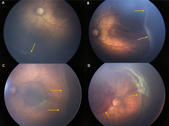
Fig. 2.

Fundus photos of stages 1–4 retinopathy of prematurity (ROP). (A) In stage 1, a demarcation line (arrow) between a normally vascularized retina and the peripheral avascular retina is shown. (B) In stage 2, the demarcation line becomes an elevated ridge (arrow). (C) In stage 3, extraretinal fibrovascular proliferation appears (arrow). (D) Partial retinal detachment (arrow) in the nasal side of the fundus and preretinal hemorrhage (dashed arrow) are shown. (Fundus photos courtesy of Dr. Sang Jin Kim).
