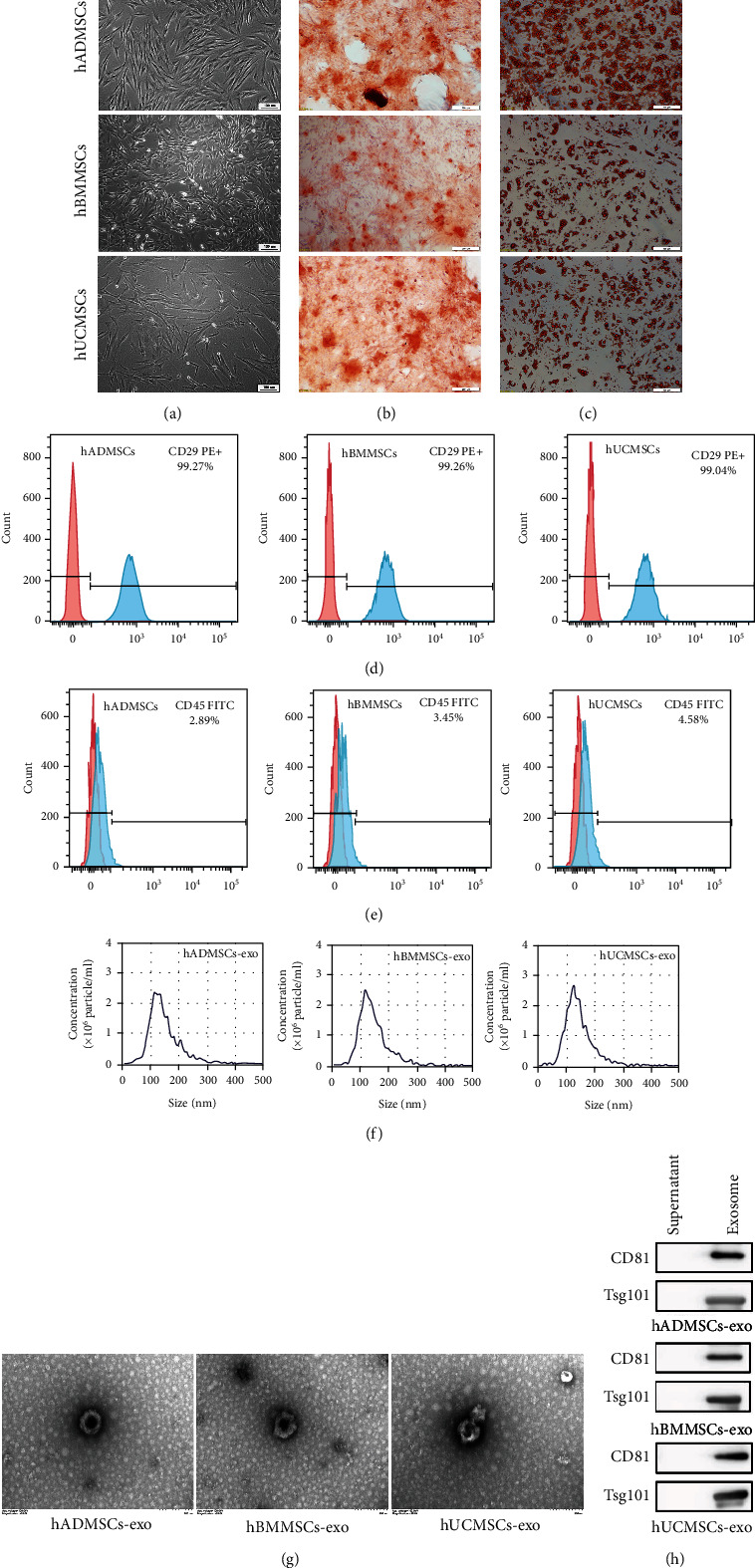
Figure 1.

Characterization of human mesenchymal stem cells (hMSCs) and hMSC-derived exosomes (hMSC-exo). (a) Morphological observation of hMSCs derived from adipose tissue (hADMSCs), bone marrow (hBMMSCs), or umbilical cord (hUCMSCs). hMSCs had a long spindle shape and were arranged in an organized fashion. Scale bar, 100 μm. (b, c) The multidifferentiation potential of hMSCs in vitro. Alizarin red S staining was used to evaluate (b) osteogenic differentiation, while oil red O staining was used to evaluate (c) adipogenic differentiation capacity. Scale bar, 100 μm. (d, e) Flow cytometric analysis of hBMSC surface markers. Note that all hMSCs were (d) positive for CD29 and (e) negative for CD45. The red shape represents the target antibody, and the blue shape represents the isotype control antibody. (f) Nanoparticle tracking assay showed the size distribution of exosomes. (g) hADMSC-exo, hBMMSC-exo, and hUCMSC-exo under the transmission electron microscope. Scale bar, 200 nm. (h) Western blot showing that exosomes derived from the three types of hMSCs were TSG101- and CD81-positive.
