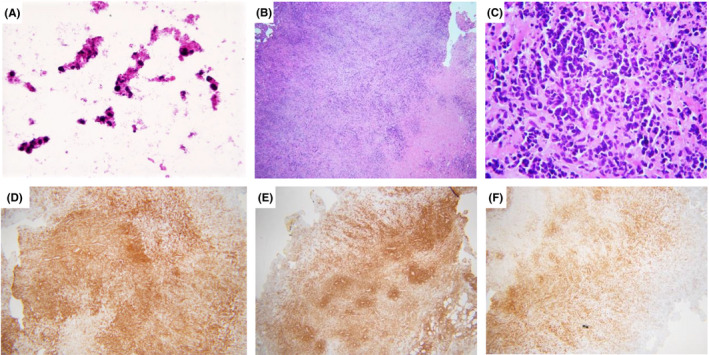
FIGURE 1.

(A) Touch imprints of retroperitoneal mass showed necrosis and undifferentiated atypical cells. (B‐C) The H&E‐stained sections of the retroperitoneal mass showed large atypical cells embedded in a dense sclerotic matrix with abundant apoptotic cells and tumor necrosis. By immunohistochemistry, neoplastic cells positive for CD45 (A), CD43 (B), and CD8 (C). Original magnifications; A: 40× B, D‐F: 4×; C: 40×
