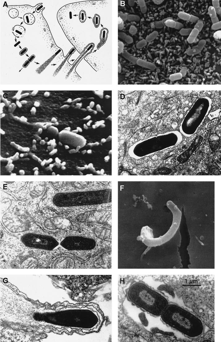
FIG. 3.
Stages of listerial intracellular parasitism. (A) Scheme of the intracellular life cycle of pathogenic Listeria spp. (reproduced from reference 655 with permission of the Rockefeller University Press). In anticlockwise order: entry into the host cell by induced phagocytosis, transient residence within a phagocytic vacuole, escape from the phagosome into the cytoplasm, cytosolic replication and recruitment of host cell actin onto the bacterial surface, actin-based motility, formation of pseudopods, phagocytosis of the pseudopods by neighboring cells, formation of a double-membrane phagosome, escape from this secondary phagosome, and reinitiation of the cycle. (B to H) Scanning and transmission electron micrographs of cell monolayers infected with L. monocytogenes (B) Numerous bacteria adhering to the microvilli of a Caco-2 cell (30 min after infection). (C) Two bacteria in the process of invasion (Caco-2 cell, 30 min postinfection). (D) Two intracellular bacteria soon after phagocytosis, still surrounded by the membranes of the phagocytic vacuole (Caco-2 cell, 1 h postinfection). (E) Intracellular Listeria cells free in the host cell cytoplasm after escape from the phagosome (Caco-2 cell, 2 h postinfection). (F) Pseudopod-like membrane protrusion induced by moving Listeria cells, with the bacterium being evident at the tip (brain microvascular endothelial cell, 4 h postinfection; taken from reference 245 with permission). (G) Section of a pseudopod-like structure in which a thin cytoplasmic extension of an infected cell is protruding into a neighboring noninfected cell (notice that the protrusion is covered by two membranes) (Caco-2 cell, 4 h postinfection). (H) Bacteria in a double-membrane vacuole formed during cell-to-cell spread (Caco-2 cell, 4 h postinfection).

