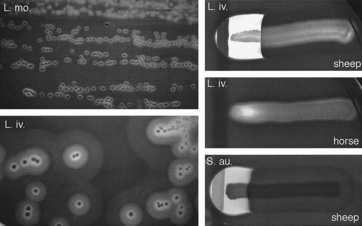
FIG. 5.
Hemolytic activities of pathogenic Listeria spp. Left panels, spontaneous hemolysis of L. monocytogenes (L. mo.) and L. ivanovii (L. iv.) on sheep blood agar; compare the narrow ring of β-hemolysis produced by L. monocytogenes (here exceptionally evident) with the striking multizonal lytic activity of L. ivanovii, which is due to the production of an additional cytolytic factor (the sphingomyelinase SmcL). Bacteria were cultured at 37°C for 36 h and then left at 4°C for 48 h. Right panels, synergistic (CAMP-like) hemolysis of L. ivanovii and β-toxin (sphingomyelinase)-producing S. aureus (S. au.) with R. equi (vertical streak); L. ivanovii and S. aureus elicit identical shovel-shaped patches of synergistic hemolysis, reflecting functional relatedness of the sphingomyelinases C produced by these two bacteria (see text and reference 226 for details). The CAMP-like effect results from the total lysis of the halo of sphingomyelinase-damaged erythrocytes exposed to a cholesterol oxidase from R. equi (467). As shown in the two upper right panels, the L. ivanovii sphingomyelinase is active towards sheep erythrocytes (which contain 51% sphingomyelin in the membrane) but not towards horse erythrocytes (13.5% sphingomyelin only). Note that the inner zone of spontaneous hemolysis that surrounds L. ivanovii colonies, due to the activity of the hemolysin (ivanolysin O), is not influenced by the source of erythrocytes. Bacteria were cultured at 37°C for 24 h.

