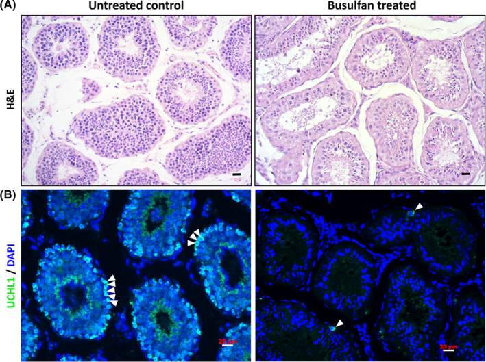
FIGURE 4.

Evaluation of seminiferous tubules in monkey testes using busulfan treatment (A) In the 60‐M‐old monkey testes, histologic analysis indicated that dramatic degeneration of spermatogenic epithelium took place in the seminiferous tubules 12 weeks after busulfan treatment (right) in comparison with the normal monkey testes (left). (B) A majority of the UCHL1‐positive spermatogonia (green) were eliminated 12 weeks after busulfan treatment (right). Cell nuclei were stained by DAPI, and staining was performed on adult rhesus testes (scale bars, 20 μm)
