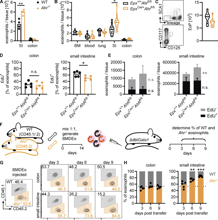
Figure 6.
AHR regulates eosinophil survival in the small intestine. (A) Eosinophils were quantified by flow cytometry in the intestine of WT and Ahr–/– mice. Data are representative of three independent experiments with n = 3–4 mice per group. **, P < 0.01, unpaired t test. (B) Eosinophil numbers (per tissue, per tibia, or per 1 ml blood) were assessed by flow cytometry across different tissues in Epx+/+Ahrfl/fl and EpxCre/+Ahrfl/fl mice. Data are pooled from three independent experiments with n = 3–7 mice per genotype. *, P < 0.05, unpaired t test with Holm–Sidak correction for multiple testing. (C) Eosinophil progenitors (EoP) in the bone marrow were identified as CD45+lineage–Sca1–CD117loCD125+ cells by flow cytometry. The number of eosinophil progenitors per tibia is shown. Data are pooled from two independent experiments with n = 3–5 mice per genotype. (D and E) Mice were treated with 1 mg/ml EdU in drinking water for 6 d. EdU+ and EdU– eosinophils in the intestine were quantified by flow cytometry. Data are representative of two independent experiments with n = 5 mice per genotype. *, P < 0.05; **, P < 0.01, unpaired t test. (F) Schematic of adoptive transfer experiments. (G and H) Percentage of WT and Ahr–/– eosinophils recovered from the colon and small intestine of recipient ΔdblGata1 mice at different time points after adoptive transfer. Dotted line, percentage of Ahr–/– eosinophils at the time of injection. Data are representative of three independent experiments with n = 3–5 mice per time point. **, P < 0.01; ***, P < 0.001, one-sample t test against the injected percentage.

