Figure S2.
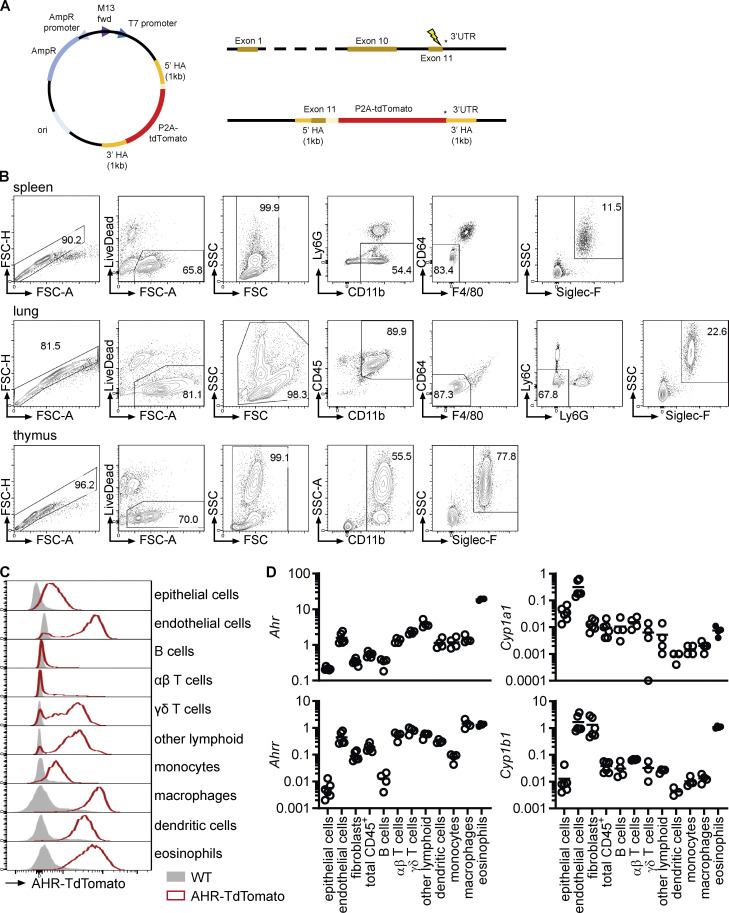
AHR expression in intestinal eosinophils. (A) Schematic of generation of AHR-TdTomato mice including DNA donor repair template, Ahr locus, and Ahr-P2A-tdTomato targeted locus. (B) Eosinophil gating and sorting strategy from different tissues for qPCR and flow cytometric analysis. Cells were positively enriched with anti-CD11b microbeads before sorting. Eosinophils from bone marrow and small intestine were sorted as shown in Fig. S1, A–C, using only LiveDead stain for dead cell exclusion. (C) AHR-TdTomato expression across different small intestinal cell types was determined by flow cytometry. Histograms are representative of n = 4 AHR-TdTomato mice from two independent experiments. (D) Gene expression was determined by qPCR across different cell types sorted from the small intestine of WT mice. Gene expression was normalized to Hprt. Data are from one experiment with four to six biological replicates. Missing data points indicate that the gene was not detectable by qPCR.

