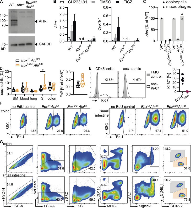
Figure S4.
AHR regulates eosinophil survival in the small intestine. (A) Western blot for AHR and GAPDH in WT, Ahr–/–, and EpxCre/+Ahrfl/fl BMDEo from culture day 14. Data are from n = 1 experiment. (B) BMDEo from culture day 14 were stimulated for 4 h with 3 μM CH223191, 5 nM FICZ, or DMSO control. Gene expression of AHR target genes was determined by qPCR and normalized to Hprt. Shown is mean ± SEM of n = 2 experiments. (C) Ahrr expression in sorted small intestinal eosinophils and macrophages of different genotypes was determined by qPCR and normalized to Hprt and is expressed as a percentage of Ahrr expression in WT for each cell type. Data are from n = 1–2 mice per genotype. (D) Frequency (percentage of CD45+ cells) of eosinophils across different tissues and of eosinophil progenitors (EoP) in the bone marrow. Data are pooled from two to three independent experiments. t test; *, P < 0.05. (E) Representative flow cytometry plots and quantification of Ki-67 staining in small intestinal eosinophils and CD45− cells (including epithelial and stromal cells). Less than 1% of eosinophils were Ki-67+. Data are from one experiment with n = 7 mice. FMO, fluorescence minus one. (F) Representative flow cytometry plots depicting EdU staining of eosinophils in the colon and small intestine after 6 d of continuous EdU administration in drinking water (1 mg/ml). (G) Representative flow cytometry plots depicting eosinophil gating in the colon and small intestine of ΔdblGata1 mice after adoptive transfer of mixed BMDEo cultures. WT and Ahr–/– eosinophils were distinguished based on their expression of CD45.1 and CD45.2.

