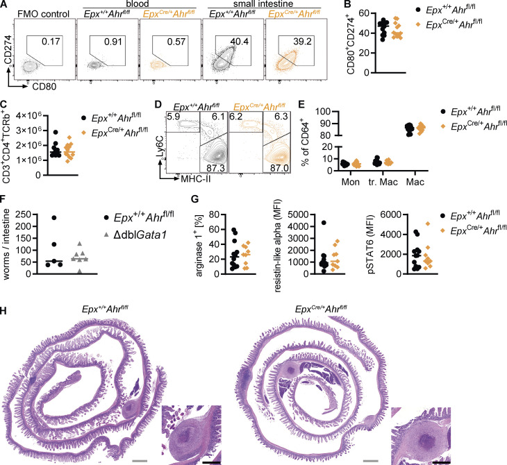
Figure S5.
AHR deficiency in eosinophils modulates the intestinal immune system. (A and B) Representative flow cytometry plots and quantification of CD80+CD274+ eosinophils in the blood and small intestine. FMO, fluorescence minus one. (C) Small intestinal CD45+CD3+CD4+TCRb+ T cells were analyzed by flow cytometry. The total cell number per small intestine is shown. (D and E) Flow cytometry of the small intestinal monocyte-macrophage compartment. Cells were pregated as live CD45+CD11b+CD64+ cells. In A–E, data are pooled from two independent experiments for a total of n = 12 mice per genotype and were analyzed by t test. (F) Worm burden was quantified in the small intestine at day 7 after H.p. infection. Data are from one experiment with n = 5–7 mice per genotype. (G) Expression of alternative activation markers in duodenal macrophages on day 14 after H.p. infection. Data are pooled from two independent experiments. MFI, mean fluorescence intensity. (H) Representative images of H&E-stained duodenum on day 28 after H.p. infection. Gray scale bars: 1 mm, black scale bars: 0.5 mm.

