Figure 1.
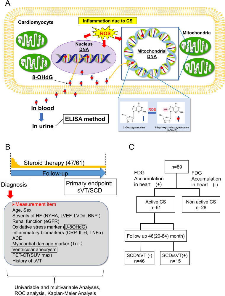
Study design. (A) 8-OHdG as a marker of DNA oxidative damage. Enhanced production of ROS may occur in cardiomyocytes during the active phase of CS, leading to DNA oxidisation (from 2’-deoxyguanosine (blue rod) to 8-OHdG (blue rod with red star)) in the nuclei DNA and mitochondrial DNA, with subsequent excretion of 8-OHdG, in urine via blood. The concentration of 8-OHdG can be measured by ELISA method using an anti-8-OHdG antibody (N45.1).10–12 The normal range of U-8-OHdG concentration is defined as <10 ng/mg·Cr, as taken from a previous study.9 (B) Study protocol. (C) Study flow chart. The 89 patients with CS were divided according to the presence of 18F-FDG accumulation in the heart into the following groups: active CS (n=61) and non-active CS (n=28). Patients with active CS were further divided into the sVT/SCD event (+) group (n=15) and the sVT/SCD event (−) group (n=46). 18F-FDG, 18F-fluorodeoxyglucose; 8-OHdG, 8-hydroxy-2′-deoxyguanosine; BNP, B-type natriuretic peptide; CRP, C reactive protein; CS, cardiac sarcoidosis; eGFR, estimated glomerular filtration rate; HF, heart failure; IL-6, interleukin 6; LVDd, left ventricular end-diastolic diameter; LVEF, left ventricular ejection fraction; NYHA, New York Heart Association; PET, positron emission tomography; ROC, receiver operating characteristic; ROS, reactive oxygen species; SCD, sudden cardiac death; SUVmax, maximum standardised uptake value; sVT, sustained ventricular tachycardia; TNF-α, tumour necrosis factor; TnT, troponin T.

