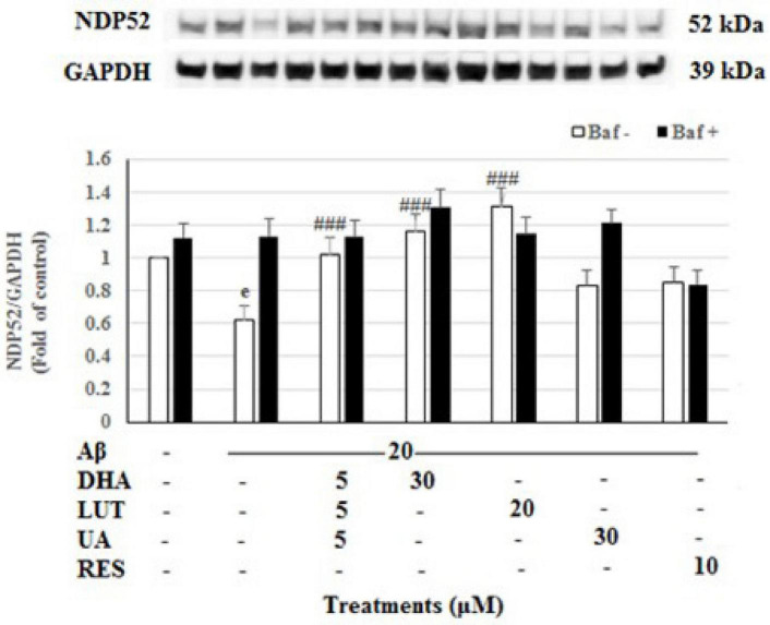
FIGURE 7.
The effect of the three-compound combination and its components on the levels of NDP52. The BE(2)-M17 cells were pre-treated for 24 h with the three-compound combination, D5L5U5, DHA (30 μM), LUT (20 μM), UA (30 μM), and RES (10 μM). The cells were then exposed to 20 μM of oligomeric Aβ1–42 for 16 h and subjected to Western blotting. The representative immunoblot and the graph indicate the levels of NDP52 protein for all treatments. Black bars indicate the NDP52 levels in the presence of bafilomycin A (Baf A) while the white bars indicate the NDP52 levels in the absence of Baf A. The relative density of the bands was normalized against GAPDH. Fold values were calculated relative to vehicle control. Data are expressed as mean ± SD from four (N = 4) independent experiments. Differences are significant at eP < 0.001 vs. vehicle control, ###P < 0.001 vs. Aβ1–42-treated controls.

