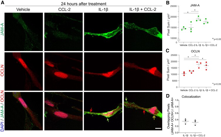
Figure 1.
Inflammation induces reactive astrocytes to express JAM-A diffusely throughout the cell surface membrane in vitro. (A and B) Astrocytic JAM-A (green) was increased in vitro at 24 h after treatment with 20 ng/ml IL-1β and, not significantly, by the combination of IL-1β + 100 ng/ml CCL-2 but not CCL-2 alone (average vehicle (14 500) versus IL-1β (18 915) versus CCL-2 (14 950) versus IL-1β + CCL-2 (17 665), vehicle versus IL-1β: P = 0.03, CCL-2 versus IL-1β: P = 0.04, one-way ANOVA with Tukey’s multiple comparison test). JAM-A was both diffusely localized throughout the cell membrane (green arrows) and co-localized with the tight junction marker, occludin (OCLN, red, red arrows; yellow double-headed arrow pointing to overlay of OCLN and JAM-A in yellow). Scale bar: 10 µm. (C) Astrocytic occludin was similarly induced at 24 h after treatment with IL-1β or the combination of IL-1β and CCL-2 but not CCL-2 alone (average vehicle (10 550) versus IL-1β (17 706) versus CCL-2 (12 117) versus IL-1β + CCL-2 (16 657), vehicle versus IL-1β: P = 0.01, vehicle versus IL-1β + CCL-2: P = 0.03, CCL-2 versus IL-1β: P = 0.04, one-way ANOVA with Tukey’s multiple comparison test). (D) The addition of CCL-2 to IL-1β did not change the proportion of JAM-A+ pixels co-localized with OCLN+ pixels at 24 h [IL-1β (0.36) versus IL-1β + CCL-2 (0.32), P = 0.63, unpaired two-tailed t-test]. (B–D) The analysis was performed on three images for each three technical replicates per condition. Dot plots show the average of the technical replicates within each biological replicate (n = 3 per group).

