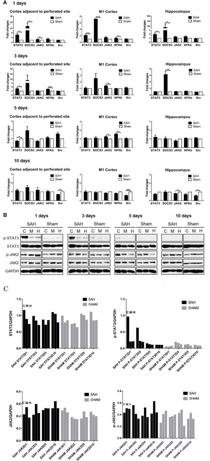
Figure 2.
STAT3 signalling pathway activated after experimental SAH. (A) RNA extract was prepared from CAPS, M1 cortex and hippocampus of SAH brain at 1, 3, 5 and 10 days. The mRNA expression of mediators involved in the STAT3 pathway after SAH was determined by RtPCR and shown in the fold change compared with the sham-operated group. n=3–4/group/time point. Values were mean±SEM. *P≤0.05, **P≤0.01, ***P≤0.001. (B) STAT3/JAK2 phosphorylation status was detected by WB at 1, 3, 5 and 10 days. The levels of phosphorylated and total STAT3/JAK2 relative to internal control GAPDH are shown in the cortex adjacent to the perforated site (C), M1 cortex (M) and hippocampus (H), respectively, of SAH and sham-operated mice. n=3/group/time point. (C) The quantitative analysis of the pSTAT3/STAT3 and pJAK2/JAK of (B) to show the trend of pSTAT3/STAT3 and pJAK2/JAK in the SAH and sham groups. CAPS, cortex adjacent to the perforated site; JAK2, Janus kinase-signal transducer 2; NF-κB, nuclear factor-kappa B; RtPCR, real-time PCR; SAH, subarachnoid haemorrhage; STAT3, signal transducer and activator of transcription 3; WB, western blot.

