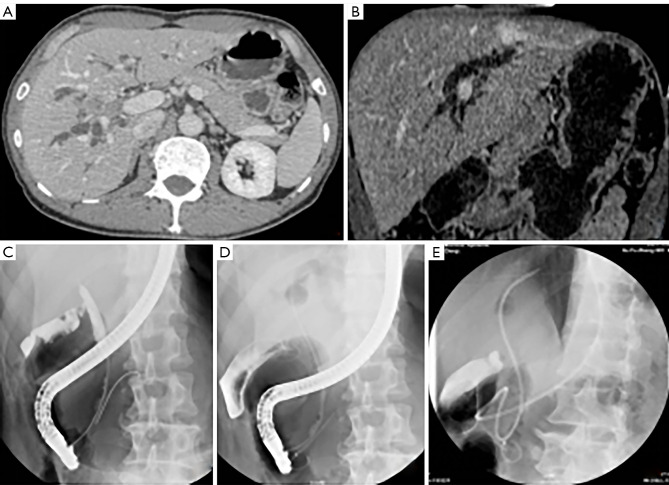
Figure 1.
Single ENBD preoperative drainage for PHC. This patient, a 66-year-old male, had Bismuth type IIIa strictures caused by hilar cholangiocarcinoma. His initial serum bilirubin level increased to 12.2 mg/dL. One 7-Fr ENBD catheter was inserted into the left lateral bile duct (future remnant lobe), and the externally drained bile was replaced by a nasointestinal tube. His serum bilirubin level decreased to 2.0 mg/dL at 42 days post-ENBD, and he had undergone segments 1, 5–8 hepatectomy. (A) Axial computed tomography with contrast shows the mass in the right liver. (B) Coronal computed tomography shows dilated left bile duct. (C) Cholangiography contrast in the common bile duct shows the distal part of the structure. (D) Air cholangiography of the left intrahepatic duct. (D) ENBD drainage for left intrahepatic duct. (E) ENBD drainage for left intrahepatic duct. ENBD, endoscopic nasobiliary drainage; PHC, perihilar cholangiocarcinoma.

