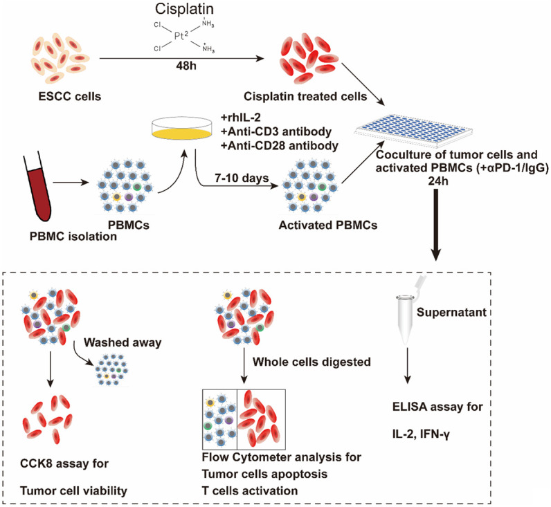
Figure 2.

Schematic diagram of co-culturing ESCC cell lines and PBMCs. K30 or TE-1 cells were cultured with CDDP (1 μg/ml) for 48 h. Thereafter, untreated or CDDP-treated K30 or TE-1 cells (5×10⁴ cells) were cultured with activated PBMCs (with 0.1 mg/ml IgG or with 0.1 mg/ml sintilimab) in 96-well round plates for 24 h, E:T=5:1. Subsequently, cell viability was detected by CCK8 assay and flow cytometry. The levels of IL-2 and IFN-γ in supernatants were measured.
