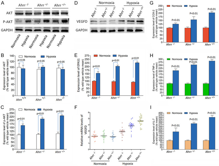
Figure 3.

Hypoxia activation of the EPAS1/PI3K/VEGFD signaling axis in Ahrr +/+, Ahrr +/- and Ahrr -/- FaDu HNC. Western blot determination of Akt (A, B) and p-Akt (C) and VEGFD (D-F) protein band density was determined using 10 µg total protein isolated from HNC cultured with vehicle or hypoxia (n=15 for each group and each Ahrr genotype). (B, C) There was no significant difference in the level of total Akt protein in the medium exposed to Ahrr +/+, Ahrr +/- and Ahrr -/- FaDu HNC, nor was there any difference with any Ahrr genotype exposed to hypoxia. (D) The increased sensitivity of Ahrr +/+ FaDu HNC to VEGFD under hypoxia cannot be explained by the increased expression of Akt protein. (E, F) These results suggest that the greater sensitivity to stimulating VEGFD production in Ahrr +/+ FaDu HNC occurs at a point downstream of Akt activation. (G-I) Under hypoxia, tumor cells release more inflammatory factors. Band density was calculated for each protein and expressed relative to band density of GAPDH (P<0.01).
