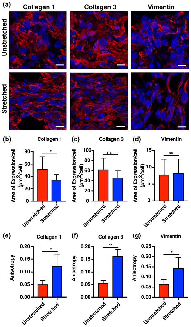
FIGURE 3.

Mechanical conditioning of engineered heart tissue grafts results in directional deposition and organization of extracellular matrix. (a) Immunofluorescent staining for collagen I, collagen III, and vimentin revealed increased directionality of extracellular matrix proteins and intermediate filament proteins in stretched compared to unstretched tissues (red = collagen I, collagen III, or vimentin, blue = DAPI; scale bar = 10 μm). (b–d) Quantification reveals increased area of expression per cell for collagen I in the unstretched compared to the stretched tissues, and no significant difference in area of expression in collagen III and vimentin between the two groups. (e–g) Anisotropy increased for collagen I and collagen III fibers and vimentin intermediate filaments in the stretched compared to the unstretched tissues (bars represent mean ± standard deviation, *p < 0.05 and **p < 0.001, n = 3) [Colour figure can be viewed at wileyonlinelibrary.com]
