
FIGURE 2.
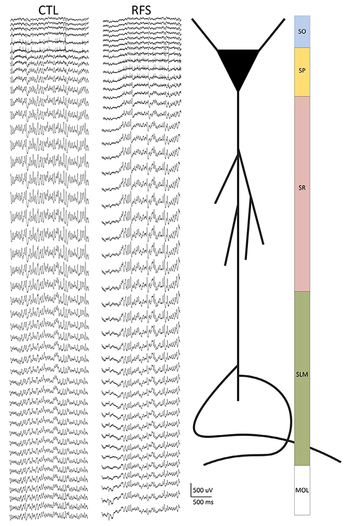
Recordings were made along the somatodendritic axis of CA1 using 64-channel laminar silicon probes in control (CTL) and experimental recurrent febrile seizure (eRFS) rats. Left: Example traces (2 s) from CTL and eRFS recordings showing the resolution of 20-μm spacing (1280 μm total) from the laminar probe. Spikes are visible on channels 5–7 (CTL) and 8–12 (eRFS) to illustrate close but variable distribution of the probe within the tissue. Stratum pyramidale (SP) was used as a reference point for probe placement during the experiment. Right: schematic of where the depth of the recording corresponds to a cartoon CA1 neuron and corresponding synaptic input regions along the somatodendritic axis. MOL, molecular layer of dentate gyrus; SLM, stratum lacunosum moleculare; SO, stratum oriens; SR, stratum radiatum