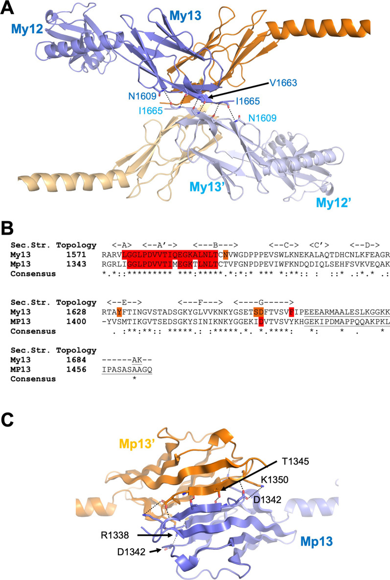
Fig. 4.
Domain 13 of myomesin-1 and -2. A Cartoon representation of the domain My13 as a tetrameric assembly highlighting the interactions of the second interface found in the crystals (PDB IDs 2R15 and 2Y25). Chain A (in blue) interacts with a space group symmetry related chain A from a neighbouring dimer. Hydrogen bonds and salt bridges are shown as dashed lines. The same interactions are detected in chain B molecules (orange and light orange). B Sequence alignment of domains My13 and Mp13. The amino-acids involved in the dimerization interface on My13 are highlighted in red as well as the conserved amino-acids of the interface on Mp13. The C-terminal amino acids of the proteins after domain 13 are underlined. C Cartoon representation of the Mp13 dimeric model. Hydrogen bonds and salt bridges are shown as dashed lines

