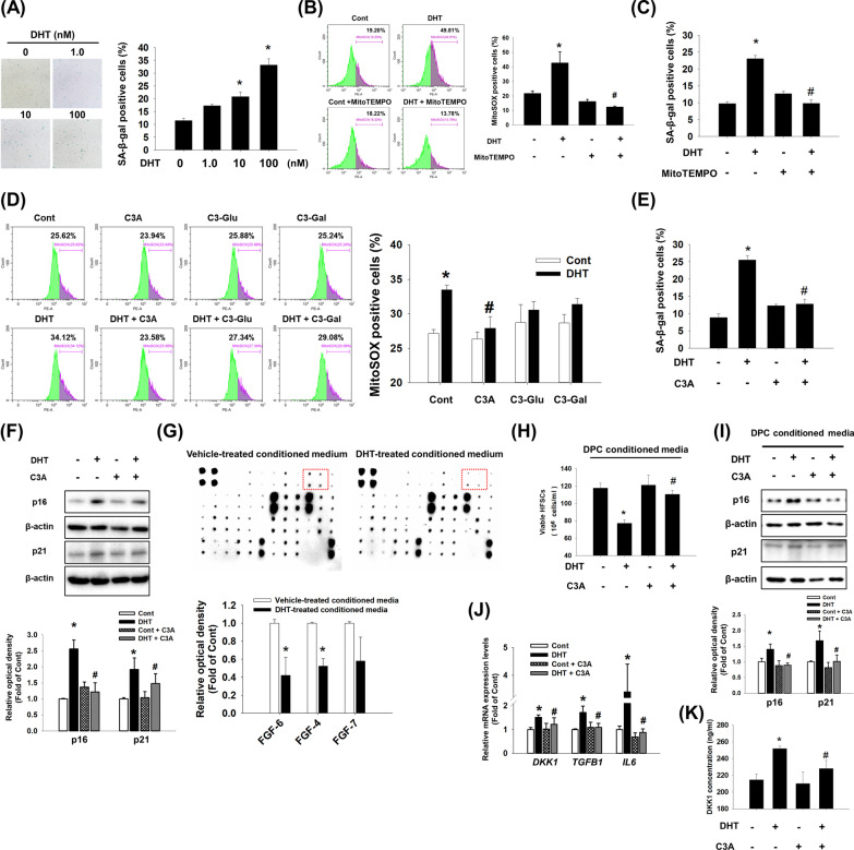
Fig. 1.
Effect of C3A on DHT-induced DPC senescence. A Dermal papilla cells (DPC) were treated with dihydrotestosterone (DHT) (0 to 100 nM) for 72 h. Senescent cells were determined by senescence associated β-galactosidase activity (SA-β-gal) assay. The positive cells (blue) were counted in five random fields manually. Proportional number of positive cells was presented by the percentage of cells of each treat group. N = 5. B Cells were pretreated with MitoTEMPO (1 μM) for 30 min and treated with DHT (100 nM) for 48 h. Cells were stained with MitoSOX and analyzed by flow cytometer. N = 4. C Cells were pretreated with MitoTEMPO (1 μM) for 30 min and treated with DHT (100 nM) for 72 h. Senescent cells were detected with SA-β-gal assay. N = 5. D Cells were pretreated with cyanidin 3-O-arabinoside (C3A) (1 μM), cyanidin-3-O-glucoside (C3-glu, 1 μM), and cyanidin-3-O-galactoside (C3-gal, 1 μM) for 30 min and treated with DHT for 48 h. Cells were stained with MitoSOX and analyzed by flow cytometer. N = 4. E Cells were pretreated with C3A for 30 min and exposed to DHT for 72 h. SA-β-gal assay was performed. N = 5. F Cells were pretreated with C3A for 30 min and treated DHT for 72 h. The protein expression levels of p16 and p21 were analyzed by western blot analysis. β-actin was used as a loading control. N = 4. G DPCs were treated with vehicle or DHT for 72 h. DPC conditioned media was used for growth factor antibody array. The results were captured by chemiluminescence imaging system and quantified by relative optical densities of spots. FGF-6, FGF-4, and FGF-7 expression levels were compared with control. N = 3. H, I DPC conditioned media were obtained from the culture media of DPCs pretreated with C3A and treated with DHT for 72 h. Human hair follicle stem cells (HFSCs) were treated with DPC conditioned media for 48 h. H Viable cells were counted with trypan blue exclusion assay. N = 6. Data are shown by cell numbers in 1 ml. I Protein expression levels of p16 and p21 were evaluated by western blot analysis. N = 4. J DPCs were treated with C3A for 30 min prior to DHT exposure for 24 h. The mRNA expression levels of DKK1, TGFB1, and IL6 analyzed by using qPCR. N = 5. K DKK-1 concentration in the DPC conditioned media by using DKK-1 ELISA kit. DPC conditioned media was taken from the culture media of DPCs pretreated with C3A and treated with DHT for 72 h. N = 5. Data are mean ± SEM. *p < 0.05 versus Control. #p < 0.05 versus DHT

