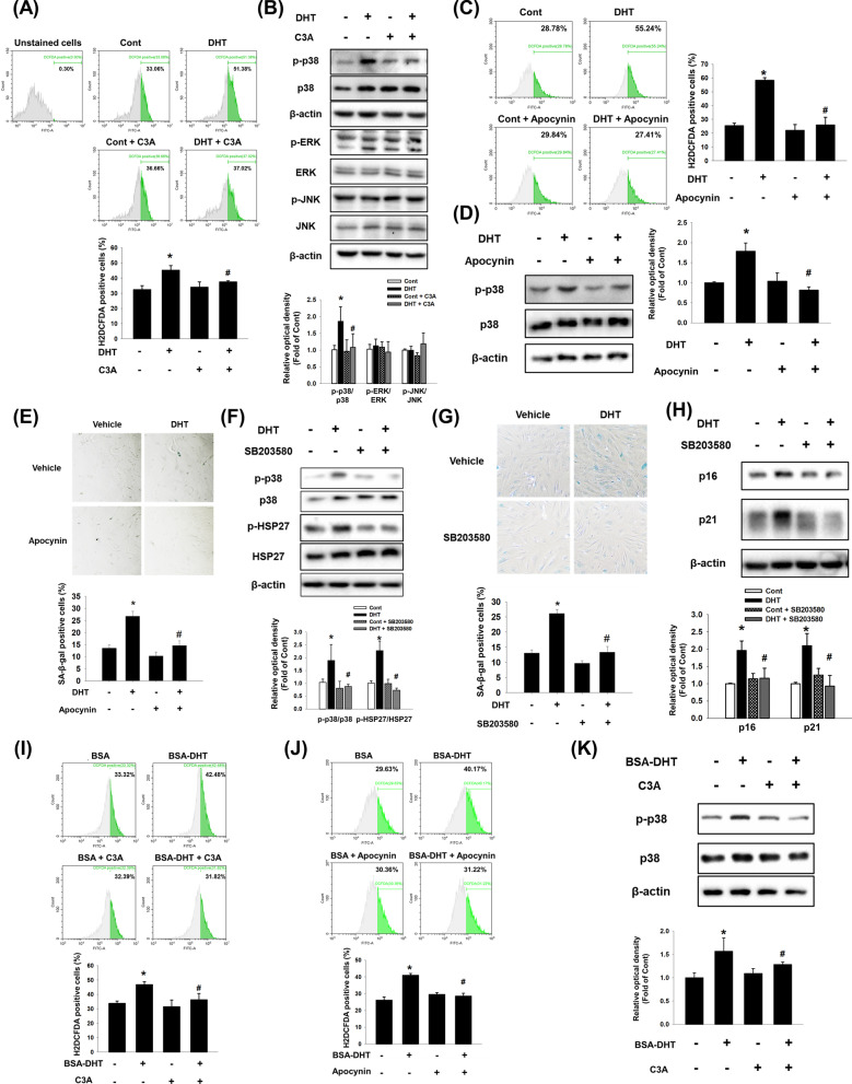
Fig. 3.
Effect of C3A on DHT-induced p38 phosphorylation. A, B DPCs were treated with C3A for 30 min and treated with DHT for 2 h. A Cells were stained with H2DCFDA (1 μM) and analyzed with flow cytometer. N = 3. B The levels of p-p38, p-ERK, and p-JNK were determined by western blot analysis. Data are normalized by each anti-p38, -ERK and -JNK antibodies. N = 3. C, D Cells were treated with Apocynin (100 μM) for 30 min and exposed to DHT for 2 h. C Cells were stained with H2DCFDA and analyzed by flow cytometer. N = 3. D Phosphorylated p38 expression level was analyzed by western blot analysis. N = 4. E Cells were treated with Apocynin for 30 min and exposed to DHT for 72 h. SA-β-gal activity assay was performed, and blue stained cells of total cells were counted. N = 5. F Cells were treated with SB203580 (1 μM) for 30 min and exposed to DHT for 24 h. Phosphorylated p38 and HSP27 expression levels were analyzed by western blot analysis. N = 3. G, H Cells were treated with SB203580 (1 μM) for 30 min and exposed to DHT for 72 h. G SA-β-gal activity assay was performed, and blue stained cells of total cells were counted. N = 5. H Western blotting was achieved for analyzing p16 and p21 protein expression levels. N = 3. I Cells were treated with C3A for 30 min and exposed to BSA conjugated DHT for 2 h. Cells were stained with H2DCFDA (1 μM) and H2DCFDA-positive cells were analyzed by flow cytometer. N = 6. J Cells were treated with Apocynin for 30 min and exposed to BSA conjugated DHT for 2 h and stained with H2DCFDA (1 μM). H2DCFDA-positive cells were analyzed by flow cytometer. N = 3. K Phosphorylated p38 expression level was analyzed by western blot. N = 4. Data are mean ± SEM. *p < 0.05 versus Control. #p < 0.05 versus DHT or BSA-DHT

