
Fig. 3.
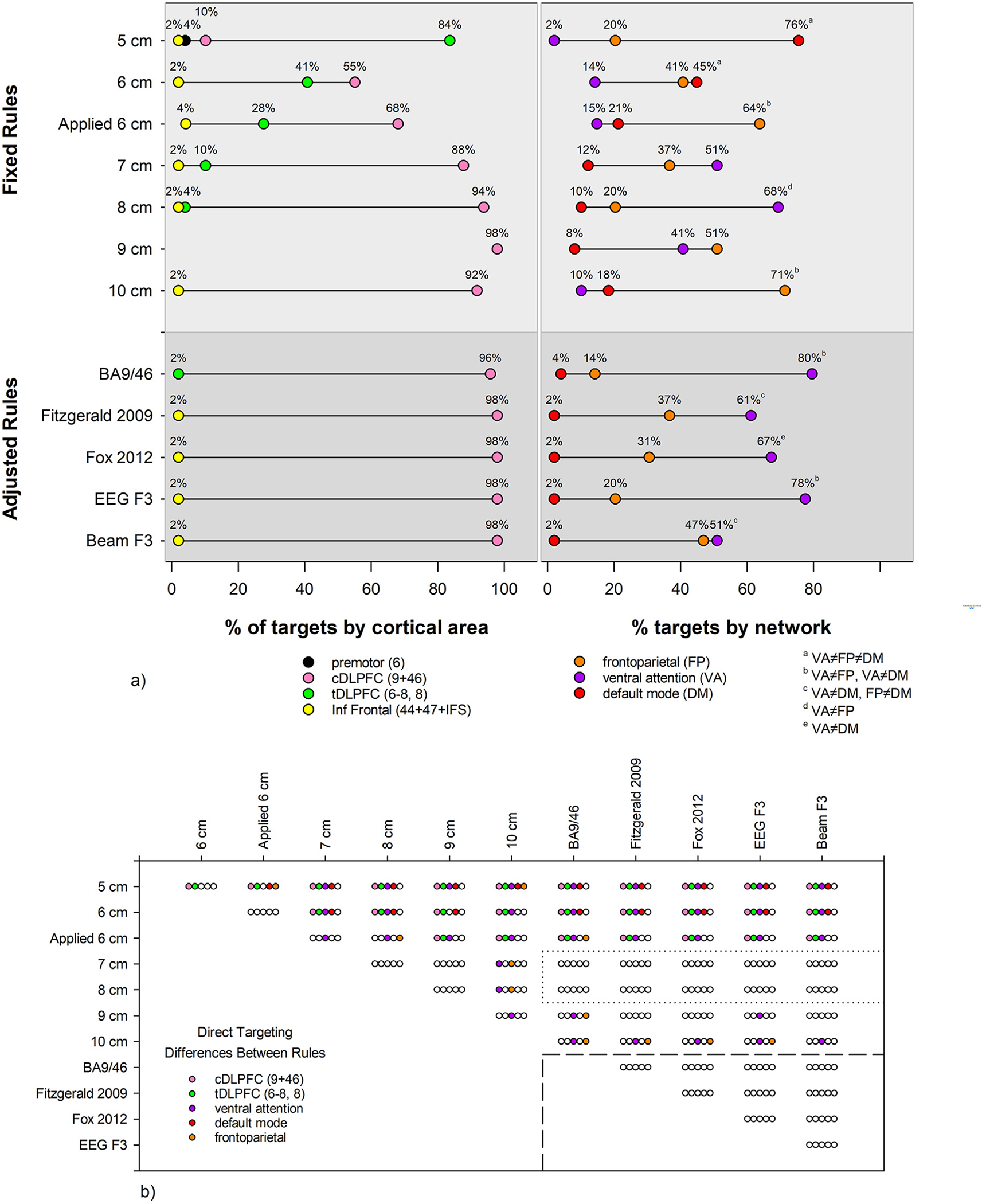
a) For each rule, the percentage of locations within HCP anatomical structure (left) and Yeo-7 network (right) is displayed. tDLPFC: transitional dorsolateral prefrontal cortex, Areas 8, 6–8; cDLPFC: classical dorsolateral prefrontal cortex, Areas 9 and 46; Inf Frontal: inferior frontal. The percentage of targets in tDLPFC and cDLPFC were different for all rules. For the % targets by network, superscript letters denote which networks were differently targeted for each rule, i.e. the 5 cm preferentially targets the default mode network, because the % of targets in the ventral attention, frontoparietal, and default mode networks are all significantly different. b) The results of χ2 tests, corrected for multiple comparisons, are shown. Each set of 5 bubbles shows whether the percentage of targets are different between the row and column rules labels, where an empty bubble means no difference. For example, the top left set of bubbles compares the 5 and 6 cm rules, with a difference in the percentage of targets in cDLPFC and tDLPFC, and no difference in networks targeted. Note that the 7, 8, and adjusted rules show no differences with each other in direct targeting of anatomy or networks.