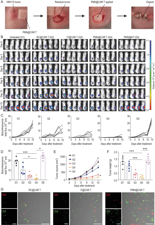
Figure 4.
CAR T cells delivered by PMN show enhanced antitumor effects in the post-surgical resection melanoma model. (A) Schematic of the mouse model in which melanoma engrafted subcutaneously is partially resected and CAR T cells delivered within the resection cavity. (B) Representative tumor bioluminescence of WM115-bearing mice treated with CSPG4+ CAR T cells administered with different modalities. Untreated mice and mice treated with a PMN loaded with control T cells were used as control. (C) Kinetics of tumor bioluminescence and (D) tumor bioluminescence at day 15 post-treatment. (E) Tumor growth curve and (F) tumor weight after treatment, n = 5. (G) Representative immunofluorescence showing CD4+ and CD8+ T cells within the tumor after treatment; scale bars: 100 μm. Data are presented as mean ± s.d., and statistical significance was calculated via one-way ANOVA with a Tukey post-hoc test. P value: *P < 0.05, ***P < 0.001.

