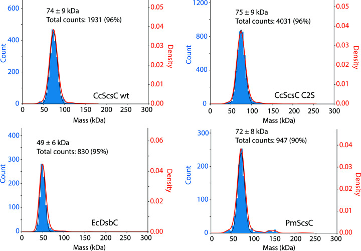
Figure 5.
Mass-photometry experiments indicate that CcScsC forms a trimer in solution. Each panel represents the mass distribution for the indicated sample (CcScsC wt and CcScsC C2S); EcDsbC and PmScsC were used as dimeric and trimeric thioredoxin-fold protein controls. Proteins were diluted to 150 nM and slowly added to reference buffer (25 mM HEPES pH 7.5, 150 mM NaCl) until around 1000 binding events per minute were observed and recorded. The mean mass of each peak, the standard deviation and the total number of events included in the peak (percentage of total events) are reported at the top of each peak. The peak positions had average masses of 75 ± 9 and 74 ± 9 kDa for CcScsC C2S and CcScsC wt, respectively, indicating a trimeric state in solution (the mass of a monomer is 25.1 kDa). Controls were included as a reference: EcDsbC showed a peak at 49 kDa corresponding to a dimer (24.1 kDa monomer) and PmScsC showed a peak at 72 kDa corresponding to a trimer (24.5 kDa monomer) consistent with previous crystal structure determinations (Furlong et al., 2017 ▸; Zapun et al., 1995 ▸).

