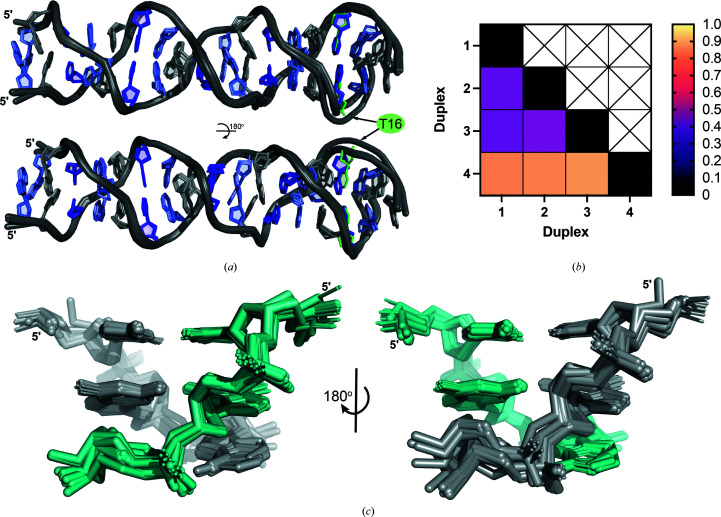
Figure 3.
d(CGA) triplets in the ps-duplex form are structurally isomorphous. (a) Overlay of duplexes 1–4 illustrating the robust structural uniformity of the ps-duplex form across different sequences, solution conditions and crystal-packing arrangements. Structural deviations are primarily observed surrounding the C16T substitution position in (CGA)5TGA. Nucleotides are colored as follows: C, purple; G, light purple; A, gray; T, green. The 5′ C–CH+ homo-base pair was omitted from (CGA)5TGA in this overlay for simplicity. (b) R.m.s.d. values from pairwise alignment of duplexes 1–4. All ps-duplexes are highly similar, with r.m.s.d. values below 1.0. (c) Overlay of all d(CGA) triplets from duplexes 1–4 rotated 180° to show the difference in deviation along the phosphate backbone from each strand (colored teal or gray). Minimal overall deviations demonstrate the high structural predictability of the ps-duplex form of the d(CGA) triplet.

