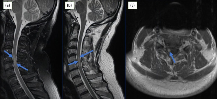
Figure 2. Cervical MRI images: (a) sagittal view demonstrating T1-weighted image with signal enhancement from C4-C6; (b) sagittal view showing T2-weighted image with hyperintense signals from C3-C5; (c) axial view of cervical spine demonstrating uniform enhancement at the level of C4.
MRI: magnetic resonance imaging, C: cervical.

