. 2022 Mar 7;11:e72416. doi: 10.7554/eLife.72416

Figure 2. Clustered protocadherins (cPcdhs) show strict homophilic specificity in their trans interactions.

(A) Surface plasmon resonance (SPR) binding profiles of cPcdh trans fragment analytes from all cPcdh subfamilies (denoted in the top row) flowed over six surfaces coated with alternate cPcdh trans fragments (rows). Responses over all surfaces are drawn on the same scale and normalized for molecular weight (mw). (B) SPR binding profiles of cPcdh trans fragment analytes from all cPcdh subfamilies (shown in columns) flowed over individual surfaces coated with C-type and α4 cPcdh trans fragments (rows). Responses over all surfaces are drawn on the same scale and normalized for molecular weight.

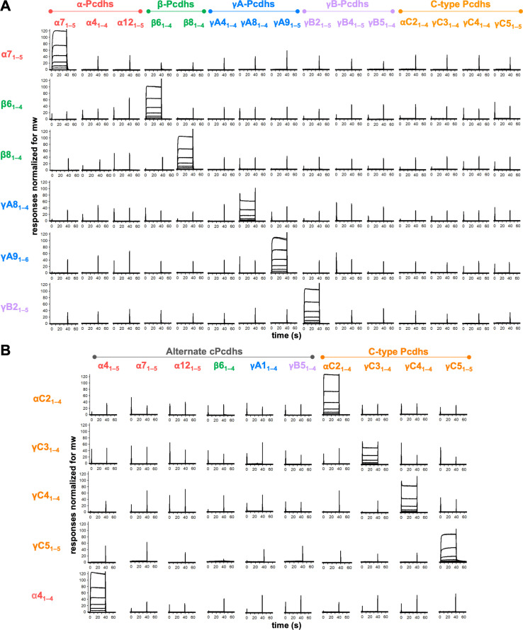
Figure 2—source data 1. Sedimentation equilibrium analytical ultracentrifugation data for trans SPR reagents.

Figure 2.

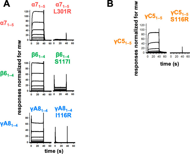
Figure 2—figure supplement 1. Trans interface mutants demonstrate homophilic interactions observed in surface plasmon resonance (SPR) are mediated by the trans dimer interface.

Figure 2—figure supplement 1.

(A) SPR-binding curves for wild-type and trans mutant alternate clustered protocadherins (cPcdhs) flowed over their respective immobilized wild-type molecule. (B) SPR-binding curves for wild-type and trans mutant C-type cPcdh γC5 flowed over immobilized wild-type γC5.
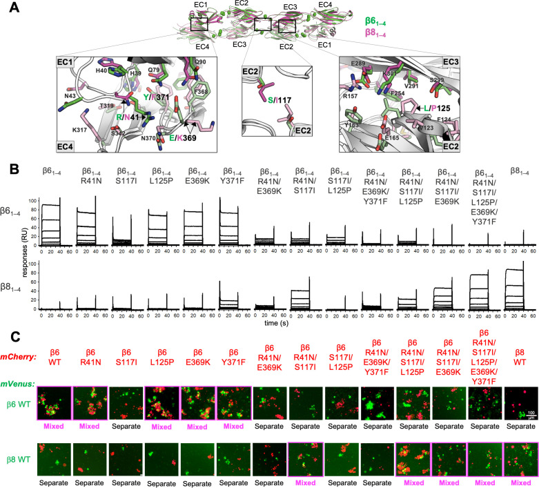
Figure 2—figure supplement 2. Mutagenesis experiments reveal role in trans specificity for the five interfacial residue differences between close pair β61–4 and β81–4.

Figure 2—figure supplement 2.

(A) Structural superposition of the β61–4 and β81–4 trans dimer crystal structures (PDBs: 5DZX and 5DZY) shown in ribbon depiction above, with close-up views of the trans interfacial regions containing the five interfacial residues that vary between β61–4 and β81–4 shown below. The two protomers forming the β61–4 dimer are colored green and pale green, respectively. The β81–4 dimer is colored magenta/light pink. Bound calcium ions are shown as green spheres. Interfacial residue side chains are shown in the close-up views. The five variable residues are labeled with the β61–4 amino acid given in green and the β81–4 amino acid in magenta: R/N41 is in EC1; E/K369 and Y/F371 are in EC4; S/I117 is in EC2 and self-interacts at the trans dimer center of symmetry; and L/P125 is also in EC2. (B) Surface plasmon resonance (SPR)-binding profiles of β6 trans interface mutants converting β61–4 to β81–4 and the wild-type molecules (shown in columns) were flowed over surfaces coated with wild-type β61–4 or β81–4 (rows). (C) Results of the K562 coaggregation assay where cells transfected with mCherry-labeled β6 and β8 wild-types (WT) and the same trans specificity mutants as in (B) were each mixed with cells transfected with mVenus labeled β6 and β8 WT. Experiments where the red and green cells coaggregate demonstrating interaction between the mCherry-labeled WT or mutant clustered protocadherin (cPcdh) and the mVenus-labeled WT cPcdh are labeled ‘mixed’ and highlighted with magenta boxes. Scale bar, 100 μM.