. 2022 Mar 7;11:e72416. doi: 10.7554/eLife.72416

Figure 5. γA4 preferentially forms the EC6-only side and γC3 the EC5–6 side in cis dimers.

(A) Structural model of γA4/γC3 cis dimer based on γB7EC3–6 cis dimer and γA4EC3–6 crystal structures (PDBs: 5V5X and 5SZQ). γA4 is shown adopting the EC6-only side (blue protomer) and γC3 is shown adopting the EC5–6 side (yellow protomer). Left, schematic of the γA4/γC3 EC3–6 cis dimer. Right, close-up view of the EC6:EC6 interface from the modeled cis dimer showing interfacial residue side chains. Bound calcium ions are shown as green spheres. Residues which were mutated in the panel B are circled in red. γB7 crystal structure numbering is used for both γA4 and γC3 residues. See methods for γA4 and γC3 alignment. Please note the model shown here is solely for hypothesis generation, since it is unlikely to be completely accurate. See methods for further details of structural modeling. (B) Top, size exclusion-coupled multiangle light scattering (SEC-MALS) data for an equimolar mixture of wild-type γA4EC3–6 and γC3EC3–6 showing dimer formation. Plot shows size exclusion absorbance at 280 nm trace (left axis), molecular weight of the eluant peaks (right axis), and the monomer molecular weights of γA4EC3–6 and γC3EC3–6 measured by mass spectrometry – 54.5 and 56.5 kDa, respectively – as dashed gray lines. Average molecular weight of the molecules in the dimer and monomer eluant peaks are labeled. Middle, SEC-MALS data for V560R mutants, which target the EC6-only side of the interface. Bottom, SEC-MALS data for residue 558 mutants. The γC3-like K558R mutation in γA4 inhibits heterodimer formation with wild-type γC3. Similarly, the γA4-like R558K in γC3 inhibits dimerization with wild-type γA4. (C) SPR-binding profiles for γB7EC3–6 wild-type and cis interface mutants flowed over three individual wild-type cis fragment surfaces. The two mutations specifically target one side of the cis interface.

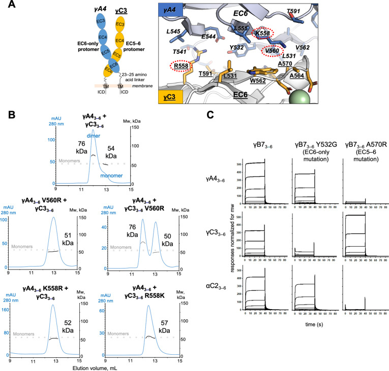
Figure 5.

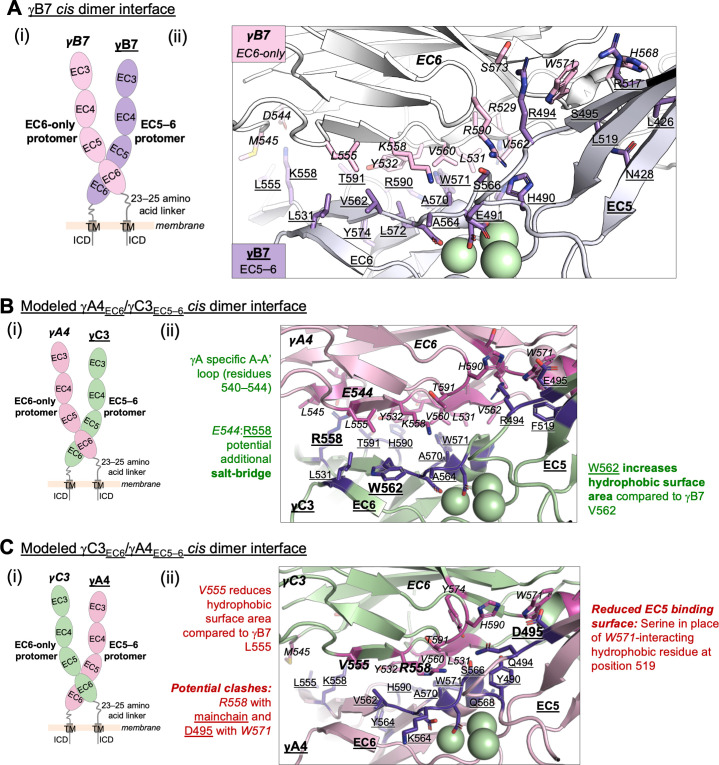
Figure 5—figure supplement 1. Structure-guided sequence analysis of γA4 and γC3 cis interactions.

Figure 5—figure supplement 1.

(A) (i) Schematic of the asymmetric γB7EC3–6 cis dimer crystal structure. (ii) Close-up view of the γB7 cis interface: interfacial residue side chains are shown in pink for the EC6-only protomer and purple for the EC5–6 protomer. Bound calcium ions are shown as green spheres. (B) (i) Schematic of the γA4EC6/γC3EC5–6 cis dimer. (ii) Model of the γA4EC6/γC3EC5–6 cis dimer interaction generated using structural alignment of EC6 from the monomeric γA4 EC3–6 crystal structure (PDB 5SZQ) to the γB7 EC3–6 cis dimer structure for the EC6-only side and computational mutagenesis of γB7 to γC3 selecting the best-fit rotamer (without energy minimization) for the EC5–6 side. The model suggests that this will be the preferred orientation for the γA4/γC3 cis dimer interaction. Favorable residue differences between γB7 from (A) and γA4/γC3 in this orientation are noted in green. Please note the model shown here is only used for hypothesis generation, since it is unlikely to be completely accurate. (C) (i) Schematic of the γC3EC6/γA4EC5–6 cis dimer. (ii) Model of the γC3EC6/γA4EC5–6 cis dimer generated using computational mutagenesis of γB7 to γC3 selecting the best-fit rotamer (without energy minimization) for the EC6-only side and structural alignment of EC5–6 from the γA4 EC3–6 crystal structure to the γB7 EC3–6 cis dimer structure for the EC5–6 side. The model suggests that this orientation for the γA4/γC3 cis dimer interaction will be disfavored. Unfavorable residue differences between γB7 and γA4/γC3 in this orientation are noted in red. Please note the model shown here is unlikely to be completely accurate and is simply for hypothesis generation.
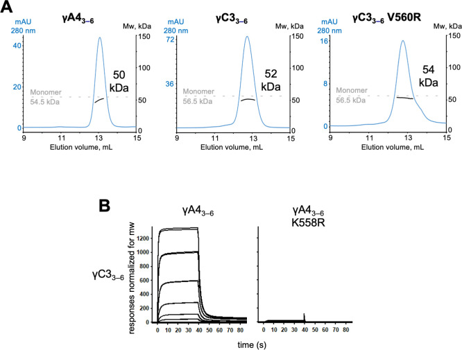
Figure 5—figure supplement 2. γA4 and γC3 cis fragments behave as monomers in size exclusion-coupled multiangle light scattering (SEC-MALS) and mutating γA4 to make it more like γC3 prevents γA4/γC3 cis heterodimerization.

Figure 5—figure supplement 2.

(A) SEC-MALS data for wild-type γA43–6, wild-type γC33–6, and γC33–6 V560R showing all three molecules are monomeric in SEC-MALS, consistent with their behavior in sedimentation equilibrium AUC. Plots show size exclusion absorbance at 280 nm trace in blue (left axis), molecular weight of the eluant peak in black (right axis), and the monomer molecular weight of γA43–6 or γC33–6 measured by mass spectrometry – 54.5 and 56.5 kDa, respectively – as dashed gray lines. Average molecular weight of the molecules in the eluant peaks are labeled. (B) Surface plasmon resonance (SPR)-binding profiles for γA43–6 wild-type and γA43–6 with γC3-like cis interface mutation K558R flowed over immobilized wild-type γC33–6. Loss of γC33–6 interaction in the presence of the K558R mutation is consistent with the SEC-MALS results shown in Figure 5.