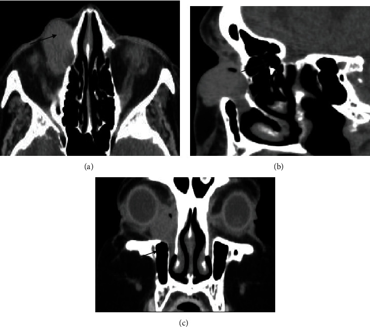
Figure 1.

(a–c) Orbital computed tomography demonstrating an oval, solid, well-circumscribed, homogeneous mass extending from the lacrimal sac into dilated duct without any evidence of invasion into adjacent bones. (c) Coronal CT scan before the second excision.
