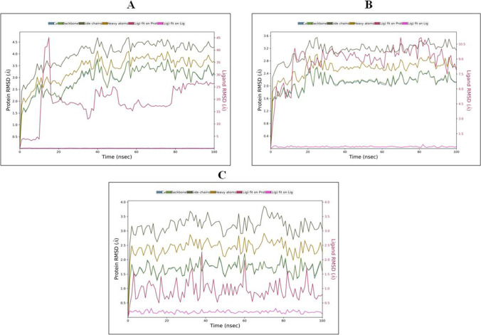
Fig. 12.
RMSD graph of best protein–ligand complexes of theophylline with SARS-CoV-2 proteins for 100 ns: (A) theophylline complex with 6LZG, (B) theophylline complex with 6LU7 and (C) theophylline complex with 6M3M. The different color lines for Cα, backbone, side chains, heavy atoms, ligand fit on protein and ligand fit on ligand are as indicated at the top of panel A, B and C

