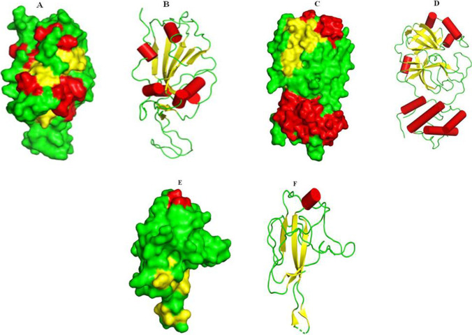
Fig. 2.
Structures of selected SARS-CoV-2 S proteins: spike protein 6LZG (A: surface view representation and B: secondary structure representation); main protease 6LU7 (C: surface view representation and D: secondary structure representation); nucleocapsid protein N-terminal RNA binding domain (E: surface view representation and F: secondary structure representation). Helices are shown in red, -sheets in yellow and loops in green color (A–F)

