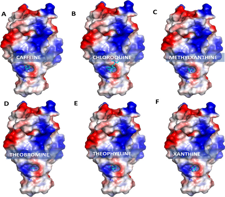
Fig. 4.

Electrostatic surface view representation of receptor-binding domain (PDB ID: 6LZG) with bound xanthine derivatives: (A) caffeine, (B) chloroquine, (C) methylxanthine, (D) theobromine, (E) theophylline and (F) xanthine. The color coding is by electrostatic potential; negative, positive and neutral regions are shown in blue, red and white respectively (image generated using Pymol)
