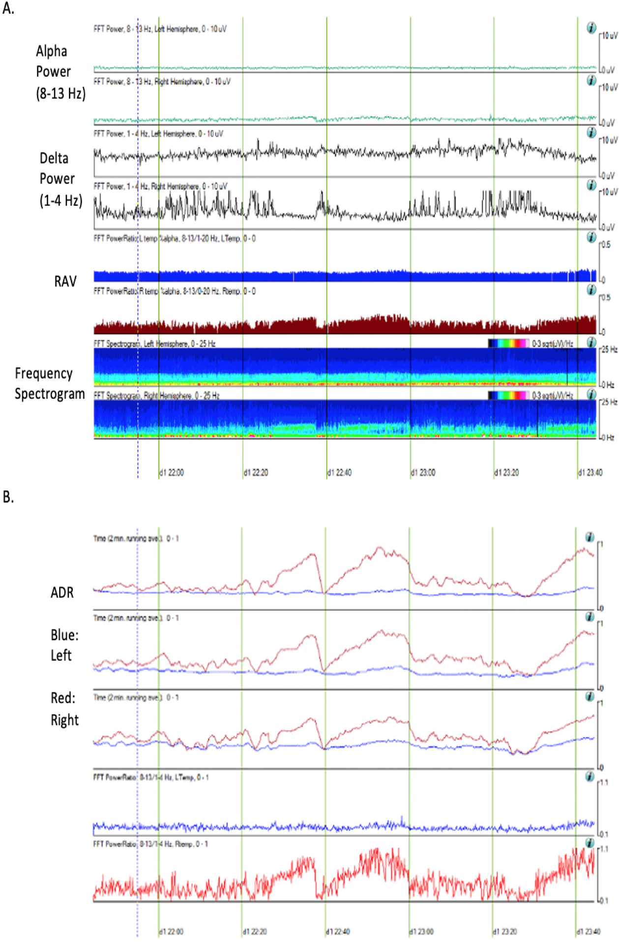
Figure 3.

qEEG parameters of a 57-year old woman at the time of DCI who presented with SAH of Hunt Hess grade 3 and modified Fisher scale 3. She developed vasospasm in the left middle meningeal artery on post-bleed day 3. The trends displayed above were generated via Persyst software version 13 (Persyst Development Corporation, Prescott, AZ). Alpha power is decreased in the left hemisphere compared to the right hemisphere while delta power is increased in the left hemisphere. The left temporal lobe shows poor relative alpha variability (RAV) but the right temporal lobe maintains good RAV (A). Alpha/delta ratio (ADR) is decreased in the left temporal lobe compared to the right temporal lobe (B).
