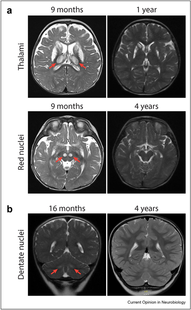
Figure 1.
T2-weighted MRI showing resolution of lesions. (a) Patient 1 basal ganglia and thalamic lesions at age 9 months with resolution of thalamic lesions at age 4 years (top) and red nuclei lesions at 9 months with resolution at age 4 years (bottom). (b) Patient 2 demonstrates improvement of dentate nuclei hyperintensities at age 16 months with marked improvement at age 4 years. MRI, magnetic resonance imaging.

