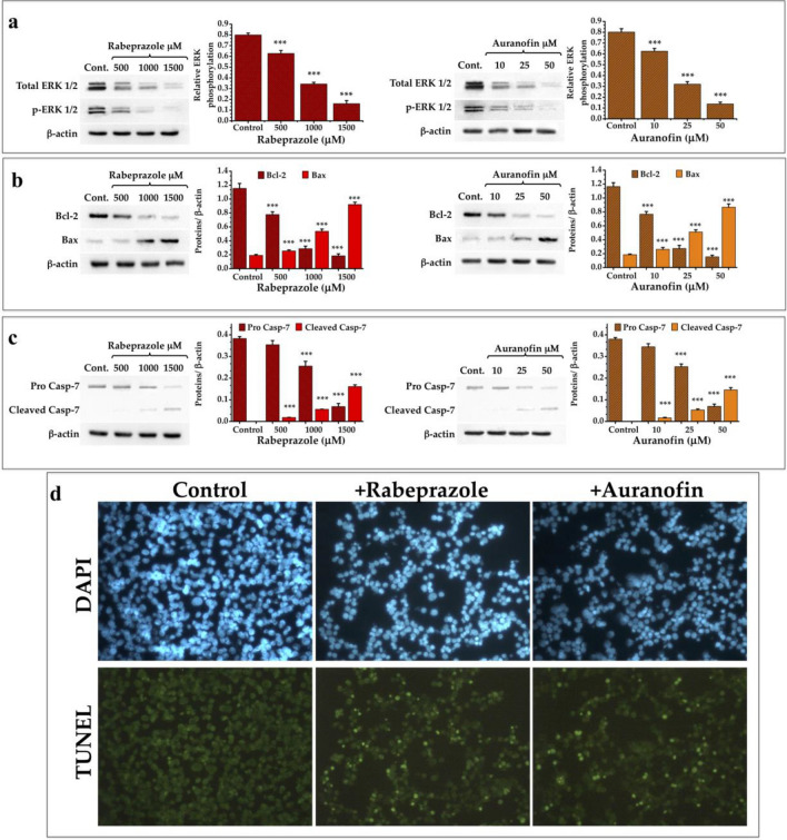
Figure 4.
Expression of elements related to programmed cell death in cancer cells treated with thiol drugs. (a) Western blot analysis of the expression of ERK 1/2 and its phosphorylated form. Graphs depict the decrease of ERK 1/2 and its phosphorylated form under treatment with rabeprazole (red bars) and auranofin (orange bars). (b) Western blot analysis of the expression of Bcl-2 and Bax. Graphs depict the expression of these two elements under treatment with rabeprazole (red bars) and auranofin (orange bars). (c) Western blot analysis of the expression of pro caspase-7 and the apparition of its cleaved form. Graphs depict the relative quantities of these two forms of Casp-7 under treatment with rabeprazole (red bars) and auranofin (orange bars). β-Actin was used as a charge control. Differences among groups were assessed with one-way ANOVA and Tukey’s test with p value = 0.001 ***. (d) TUNEL assays of cancer cells do not treated (Control) or treated with 700 μM rabeprazole (+ Rabeprazole) or 70 μM auranofin (+ Auranofin). DAPI was used for nuclei staining. Photographs are at ×40. Full-length blots of (a–c) panels are shown in Suppl. Fig. S5.

