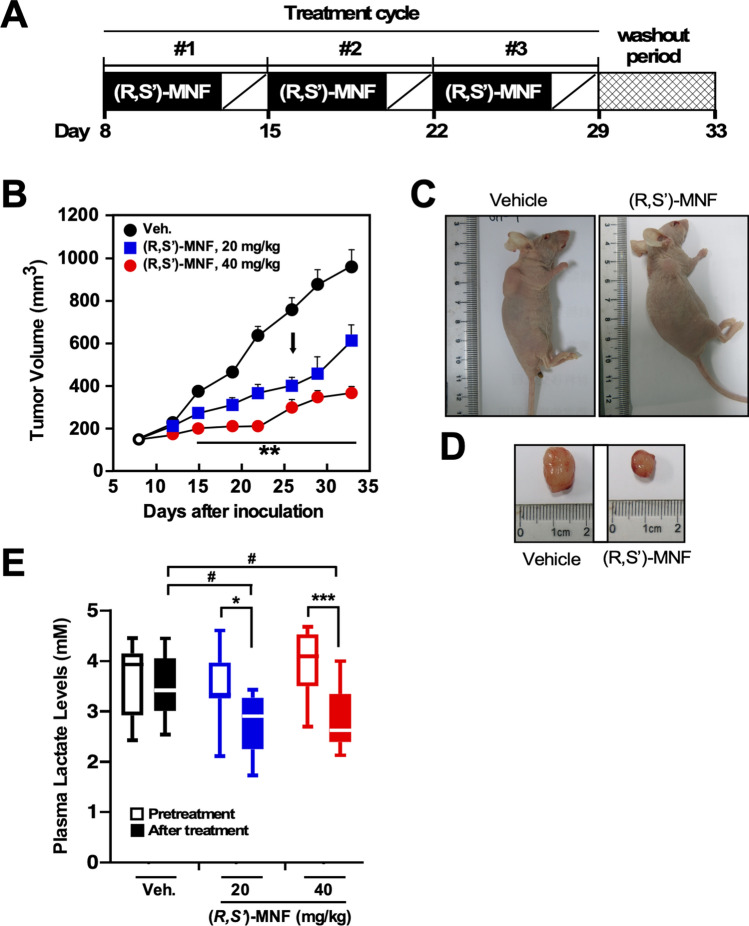
Figure 3.
(R,S′)-MNF treatment reduces PANC-1 tumor growth in a mouse xenograft model. (A) Protocol design. (B) Tumor volume was determined after i.p. administration of vehicle (1% hydroxypropyl-β-cyclodextrin), 20 mg kg−1 (Arm 1) or 40 mg kg−1 (R,S′)-MNF (Arm 2) once daily for 5 days per week for 3 weeks (n = 10 per group). The black arrow depicts the last day of (R,S′)-MNF administration. Data represent mean + SD. **P < 0.01 vs. control group of mice. Representative images of mice (C) and excised tumors (D) are shown. (E) Plasma lactate levels were measured on Day 8 (Pretreatment, open symbols) and at completion of the (R,S′)-MNF treatment (After treatment, filled symbols) (n = 8–10 per group). *P < 0.05, ***P < 0.001; #P < 0.05 vs. control group of mice at the completion of the study. The dose- and time-dependent trajectories as well as the box plot were generated with GraphPad Prism v.8.4.3 (GraphPad Software, Inc., La Jolla, CA).

