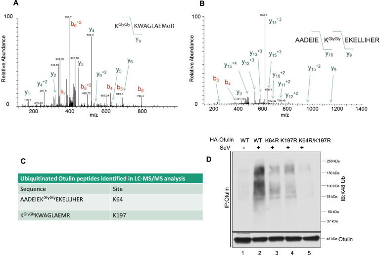
Fig. 6. Identification of the K48-ubiquitination sites.
A, B HT1080 cells were transfected with Otulin-GFP overnight followed by SeV infection for 12 h. MG132 was given for 4 h before harvesting cells. Otulin was purified with GFP-Trap beads, digested with trypsin and ubiquitinated peptides corresponding to (A) KGlyGlyKWAGLAEMR and (B) AADEIEKGlyGlyEKELLIHER and were detected by LC-MS/MS analysis. C Table showing peptides detected in LC-MS/MS analysis and the positions of lysine residues. D K48-Ubiquitination assay of Otulin WT and Otulin lysine mutants in reconstituted OT-KD cells to assess of the effect of lysine mutations on Otulin K48 ubiquitination post-SeV infection of 24 h. Immunoprecipitation assay was performed with Otulin antibody and immunoblot analysis was done with K48-Ub antibody. Quantification of western blots and statistical analyses are provided in Supplementary Fig. 8 (mean ± SEM, n = 3; ns > 0.05; *P < 0.05; **P < 0.01, ***P < 0.001).

