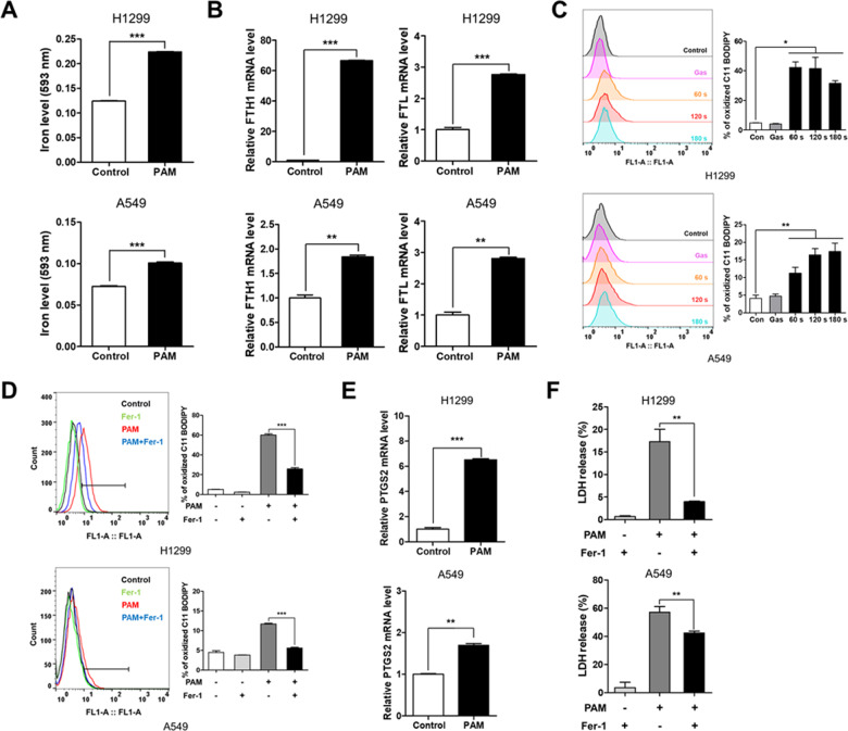
Fig. 3. Plasma-activated medium induces ferroptosis in human lung cancer cells.
The plasma exposure conditions were 1.9 standard liter per minute (SLM), 7 W input power and three exposure times (60, 120, and 180 s). A The expression levels of labile iron were measured using iron assay kit. Indicated cells were treated with PAM (180 s) for 24 h. B mRNA expression of FTH1 and FTL was measured by qRT-PCR. C PAM-induced lipid ROS levels as assessed by flow cytometry using BODIPY 581/591 C11 dye. H1299 and A549 cells were treated with PAM (180 s) for 2 h. D Lipid ROS levels were detected using BODIPY 581/591 C11 dye. The indicated cells were treated PAM (180 s) or 2 μM ferrostatin-1 for 2 h. E The expression of PTGS2, the representative ferroptosis marker, was measured by qRT-PCR. F H1299 and A549 cells were treated with PAM (180 s) or 2 μM ferrostatin-1 for 24 h. Release of LDH levels was measured using cytotoxicity kit. Data represent the mean ± SD from three independent experiments (*p < 0.05, **p < 0.01 and ***p < 0.001, as determined by t test).

