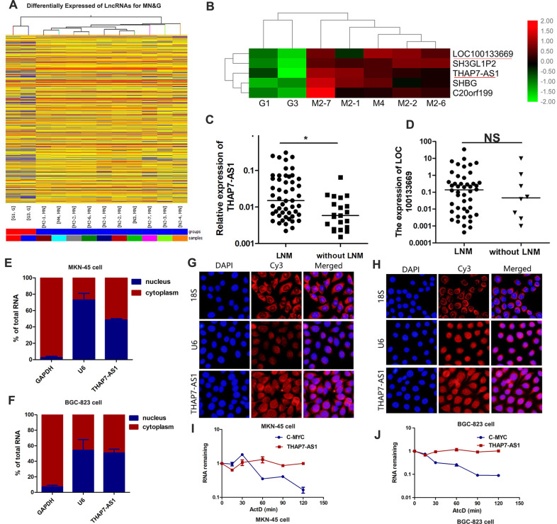
Fig. 1. THAP7-AS1 is upregulated in GC samples with lymph node metastasis.
A, B Hierarchical clustering of differentially lncRNAs expression in 10 GC cases and 2 non-tumorous gastric tissues. Each row represents different group and each column represents the expression level of an individual lncRNA. Red represents upregulated genes, and green represents down-regulated genes. C THAP7-AS1 expression was detected in human GC tissues with lymph node metastasis (LNM) and without LNM. D LOC100133669 expression was detected in human GC tissues with lymph node metastasis (LNM) and without LNM. E–H Nuclear/cytoplasm fractionation (E–F) and RNA FISH assay (G–H, ×200) were performed to observe the cellular location of THAP7-AS1 in MKN-45 cells (E, G) and BGC-823 cells (F, H). I–J RNA Polymerase II Inhibitor Actinomycin D (ActD) Chase assay was used to observe the RNA stability of THAP7-AS1 in MKN-45 cells (I) and BGC-823 cells (J), with c-Myc as a positive control for short RNA half-life in GC cells. *P < 0.05, **P < 0.01, ***P < 0.001.

