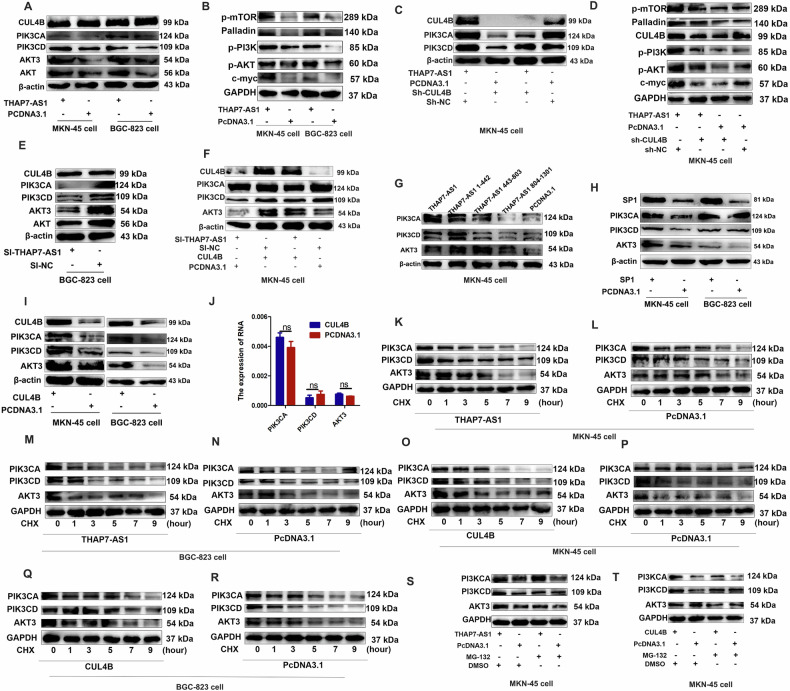
Fig. 5. THAP7-AS1/CUL4B complex initiates PI3K/AKT signaling.
A–B Western blot analysis of the different signaling modules downstream of THAP7-AS1 in both GC cells expressing THAP7-AS1 or control cells. C–D Western blot analysis of the PI3K/AKT signaling downstream of THAP7-AS1 in GC cells expressing THAP7-AS1 or control cells with or without CUL4B knockdown. E Western blot analysis of the different signaling modules downstream of THAP7-AS1 in GC cells expressing si-THAP7-AS1 or control cells. F Western blot analysis of the PI3K/AKT signaling module downstream of THAP7-AS1 in MKN-45 cells expressing si-THAP7-AS1 or control cells with or without transient transfection with PCDNA3.1-CUL4B. G Western blot analysis of the THAP7-AS1 fragments mediated downstream PI3K/AKT signaling of THAP7-AS1/ CUL4B in MKN-45 cells. H Western blot analysis of the different signaling downstream of THAP7-AS1 in both GC cells expressing SP1 or control cells. I Western blot analysis of the different signaling downstream of THAP7-AS1 in both GC cells expressing CUL4B or control cells. J The mRNA expression of PIK3CD, PIK3CA and AKT3 was detected by RT-qPCR assays. K–R MKN-45 cells (K–L and O–P) and BGC-823 cells (M–N and Q–R) transfected with THAP7-AS1(K, M) or CUL4B (O, Q) overexpression vector and control cells were treated with cycloheximide (CHX; 5 mg/mL) or vehicle for the indicated periods. PIK3CA, PIK3CD and AKT3 protein levels were analyzed by Western blotting. S–T MKN-45 cells transfected with THAP7-AS1 (S) or CUL4B (T) overexpression vector and control cells were treated with MG132 (5 mmol/L) or vehicle for 24 h. Cell lysates were analyzed by Western blotting.

