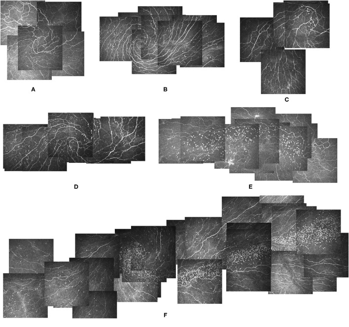
Figure 2.
Different types of globular cells distribution patterns in the corneal vortex. (A) Image from a 34-year-old male participant showing a Non-globular cell pattern and a clear vortex with no globular cells. He had no LCs. (B) Image from a 29-year-old male participant showing a Non-globular cell pattern with LCs. (C,D) Images from a 36-year-old female participant showing a type 1 globular cells distribution pattern, scattered globular cells around the vortex area with LCs (D) and without LCs 2 months after treatment (C). (E) Image from the right eye of a 57-year-old female participant showing a type 2 globular cells distribution pattern, with a large number of globular cells gathering within the vortex and distributing along the nerve horizontally in the temporal direction. She had no LCs in the vortex. (F) Image from the left eye of a 41-year-old female participant showing a type 2 globular cells distribution pattern with LCs in the vortex. The globular cells were distributed horizontally along the nerve in the temporal direction. LCs: Langerhans cells.

