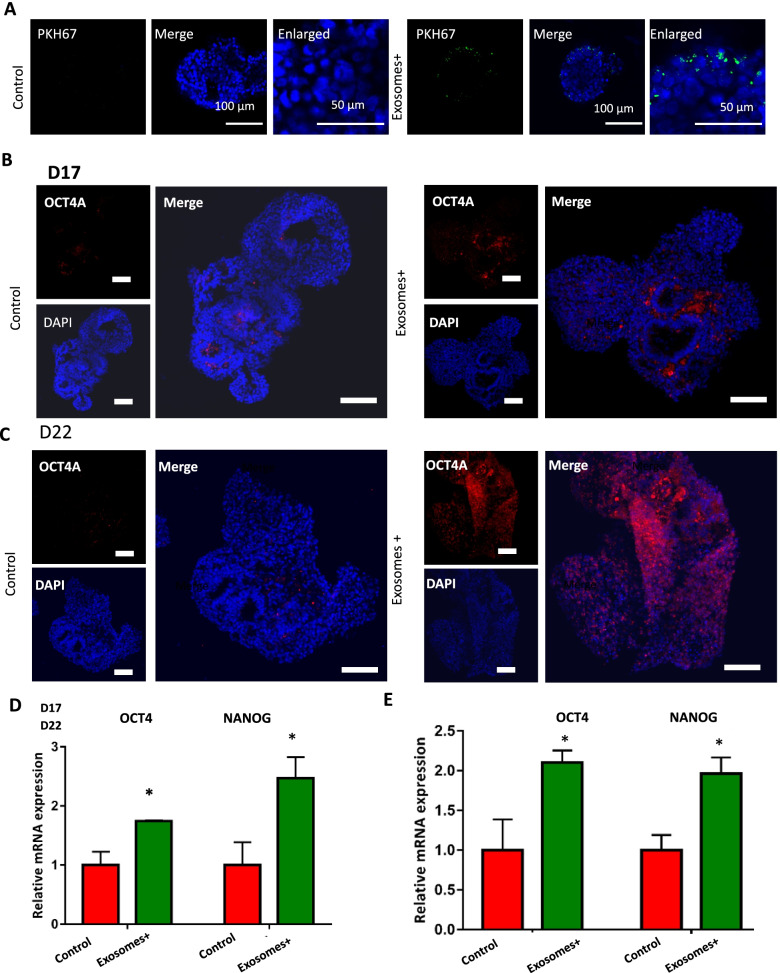
Fig. 3.
The impact of exosomes derived from breast cancer cell on the stemness of brain organoids. A Immunostaining images of cell membrane dyes PKH67 which used to mark exosomes in the developing brain organoids with and without exosome exposure. Scale bars:100 μm in the merge images and 50 μm in the enlarged images. B, C The Immunohistochemistry images of stemness marker: OCT4 in brain organoids with and without exosome exposure at day 17 and day 22. Scale bars: 100 μm. D, E The mRNA expression of stemness markers OCT4 and NANOG identified by qRT-PCR in brain organoids. Data are mean ± SEM. Student’s t-test, *P < 0.05

