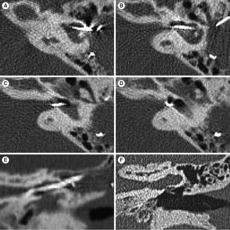
Fig. 3.

Computed tomography images of patient 3. (A-D) Axial images confirmed that the electrode did not come into contact with the inner wall of the cavity. (E) Coronal image of the straightly positioned electrode. (F) Coronal image of the same plane before surgery.
