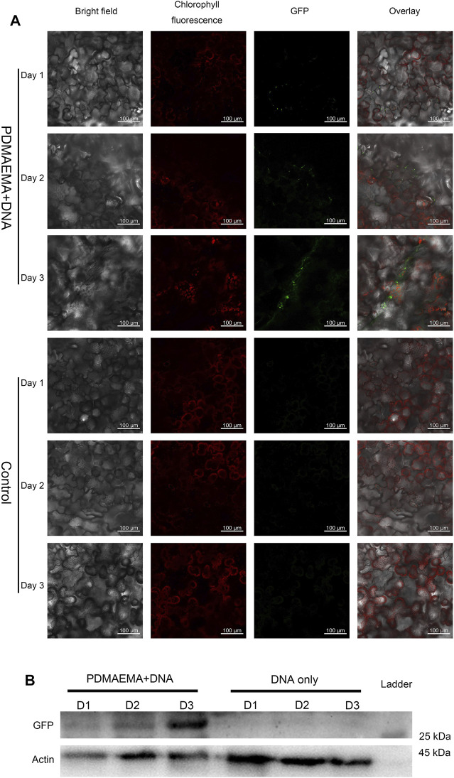
FIGURE 4.
GFP expression in Nicotiana benthamiana leaves. (A) Nicotiana benthamiana leaves infiltrated with DNA only (in 10 mM MgCl2/MES as a control) and PDMAEMA + DNA (N/P ratio of 15:1) are imaged using a confocal microscope to detect GFP expression in the leaf lamina in 1 day, 2 days, and 3 days. Experiments were performed with intact leaves from healthy plants. Scale bar, 100 μm. (B) Western blot of GFP expression of Nicotiana benthamiana leaves infiltrated with DNA only (in 10 mM MgCl2/MES as control) and PDMAEMA + DNA in 1 day, 2 days, and 3 days.

