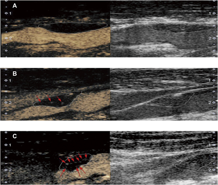
FIGURE 2.
Typical examples of plaque based on contrast-enhanced ultrasonography (left) and carotid ultrasound (right). (A) grade 0: no enhancing microbubbles within the plaque; (B) grade 1: moderate enhancing microbubbles on the shoulder and/or adventitial side of the plaque; (C) grade 2: extensive enhancing microbubbles throughout the plaque.

