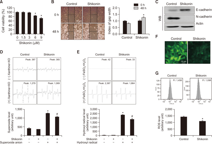
Fig. 5.
Shikonin attenuated the epithelial-mesenchymal transition in SNU-C5RR colon cancer cells. (A) SNU-C5RR cells were treated with shikonin at concentrations 0, 1.5, 3, 6, or 9 μM for 48 h, and cell viability was assessed using the MTT assay. *p<0.05 vs control cells. (B) Cells were seeded in 100 mm culture dishes, and following treatment with 3 µM shikonin, a scratch was established along the base of the dish. The wounded area was observed following treatment with or without 3 µM shikonin after 0 and 48 h. The gap was measured, and all data were quantified. Representative images for 48 h incubation are shown. *p<0.05 vs control cells. (C) Cell lysates were electrophoresed and used for western blotting with anti-E-cadherin and anti-N-cadherin antibodies. Actin was used as the loading control. The ability of 3 µM shikonin to scavenge ROS in cell-free conditions was evaluated using (D) the xanthine/xanthine oxidase system for superoxide anion and (E) Fenton reaction (FeSO4+H2O2) system for hydroxyl radical. *p<0.05 vs control, #p<0.05 vs radical only. SNU-C5RR cells were dyed with DCF-DA, and the fluorescence intensity was detected via (F) confocal microscopy, and (G) flow cytometry. *p<0.05 vs control.

