
Figure 5.
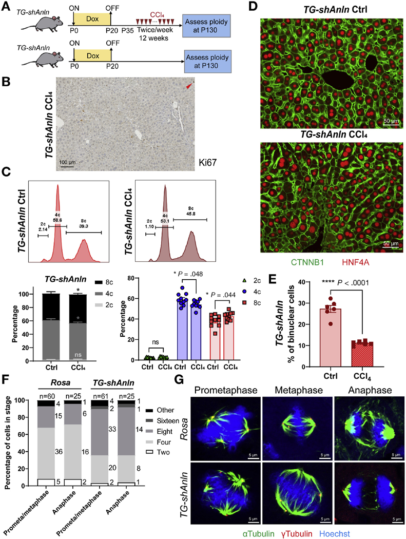
Polyploid hepatocytes in vivo readily divide and regenerate without leading to chromosome missegregation. (A) Schema for chronic CCl4 injury experiment. Doxycycline treatment established polyploidy. Twelve weeks of biweekly CCl4 started at P35. When mice were euthanized, hepatocytes were dissociated for ploidy analysis or livers were harvested for image analysis. (B) Ki-67 staining of TG-shAnln livers collected 2 weeks after the last dose of CCl4. (C) Representative cellular ploidy distribution within livers, as determined by PI staining and flow cytometry (upper). Average cellular ploidy distribution (lower left). The percentage of each population, separated from the left (right, n = 10 mice in each group were analyzed). (D) CTNNB1 (green) and HNF4A (red) stained liver sections allow ploidy analysis. (E) The percentage of binuclear hepatocytes was quantified and averaged from 4 images (n = 6 mice in each group). (F) Ploidy of hepatocytes within tissue sections of regenerating livers are estimated by centrosome number (quantified γ-tubulin foci). These hepatocytes are in prometa/metaphase and anaphase, and more than 50 and 25 mitoses in prometa/metaphase and anaphase were measured. (G) Representative images of hepatocytes within Rosa and TG-shAnln liver sections at different stages of mitosis, as stained by α-tubulin (green), γ-tubulin (red), and Hoechst (blue).