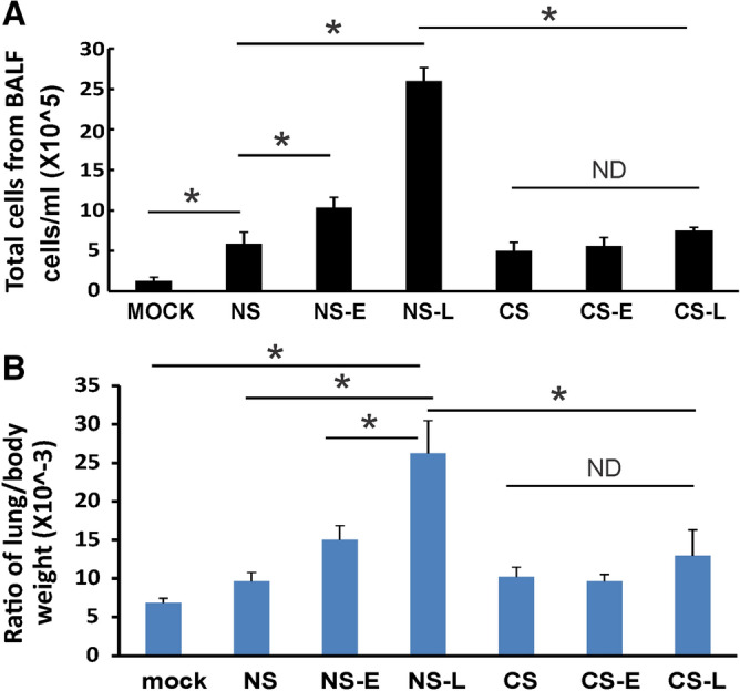
Figure 3.

Lung injury and cellularity in the lungs. CS exposure, early or late IFN-β administration and IAV infection are the same as Fig. 2A. Each mouse was infected with 500 PFU of IAV. Mock treated mice were inoculated with PBS. Bronchoalveolar lavage fluid (BALF) or lung tissue was harvested at day 5 after infection. Total immune cells (A) in BALF and ratio of lung/body weight (B) were determined. Data are expressed as means ± SEM (n ≥ 4/group). *denotes significant difference between the two groups, p < 0.05. ND = no significant difference between the two groups.
